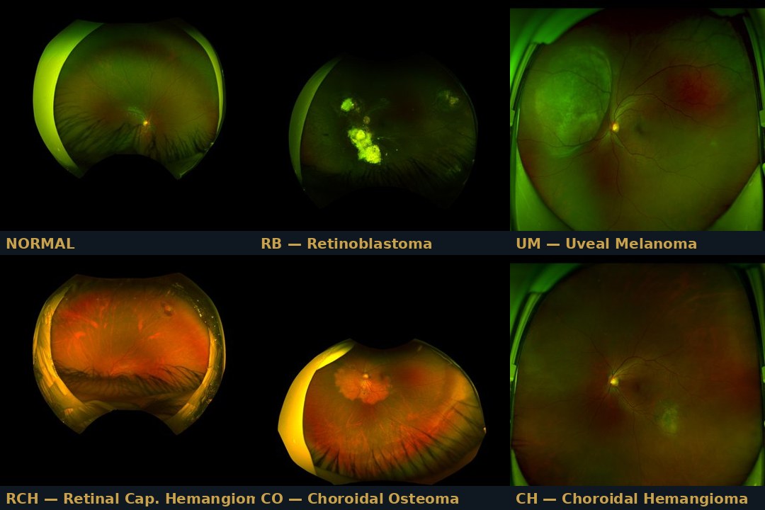
OcuGuard classifies colour fundus photographs across 6 intraocular disease classes using a 13-engine ensemble and routes every case to one of four triage zones by clinical severity — from non-diseased to immediate specialist referral. An independent out-of-distribution safety gate catches anything that is not a genuine fundus photograph before it can be classified at all. An eye-care professional always stays in command. Seven annotated demo cases span the full spectrum from a healthy fundus to the out-of-distribution safety gate itself.
Modality
Colour fundus photography · ResNet-50 transfer learning · 6 intraocular disease classes · ultra-widefield fundus research dataset
A severity-zoned triage aid for fundus review. Independent out-of-distribution gate defers anything that is not a genuine fundus photograph. Honest score, not a verdict.
🔬
Researchers
Validated ensemble metrics reported out-of-fold, per class. Honest per-class recall reported as measured. ResNet-50 transfer learning pipeline with a 13-engine ensemble. Full methodology, no cherry-picking.
🎓
Educators
A live classroom tool: seven annotated fundus cases across 6 disease classes, severity zones, and an out-of-distribution gate demonstration. Companion textbook and lab sheet for biomedical AI courses.
📖
Students
Explore how fundus features translate to AI classification. See how an ensemble routes cases by severity and why an eye-care professional must always review the result.
🏥
Hospital Planners
A demonstration of AI-augmented fundus triage. PDPA-compliant. Zone protocol ensures every Tumor, Cancer, and out-of-distribution case reaches a specialist, never a silent algorithmic verdict.
Three integrated capabilities
OcuGuard combines severity-based zone triage with an independent out-of-distribution safety gate and a six-class fundus classification engine. Each capability is designed to work together — classify the fundus image, catch anything that should never have been classified at all, and know exactly when to defer.
Capability 1
🚥 Severity-Based Zone Triage
Every fundus case is routed to one of four triage zones based on the clinical severity of the predicted class itself — not the model’s confidence in it. Clinicians act on the colour, not a percentage figure.
GREEN: Normal — non-diseased · YELLOW: Tumor group (RCH / CO / CH) — potential, advise referral to consultant · ORANGE: Cancer group (RB / UM) — strongly advised to refer to consultant · RED: Unknown — refer to consultant immediately
This is a deliberate clinical design choice: the zone reflects what the class means for the patient, so a clinician never has to translate a confidence percentage into an urgency decision themselves.
4 zones · Severity-based, not confidence-based · Specialist referral
Capability 2
⚠️ Independent Out-of-Distribution Safety Gate
Before any classification is offered, the system checks whether the submitted image actually resembles a fundus photograph at all. Anything that does not — a photo of the wrong body part, a non-medical image, a corrupted upload — is flagged Unknown and routed to the most urgent zone, RED, rather than forced into one of the six disease classes.
This means the system never guesses a diagnosis out of an image it was never designed to read. An unclassifiable image is treated as the highest-uncertainty case, not the least informative one.
The gate operates independently of the 6-class classifier — it decides whether to classify at all, before the classifier ever runs.
Fires before classification · Routed to RED · Always disclosed
Capability 3
📊 6-Class Fundus Classification with Subtype Detail
The 13-engine ensemble classifies across Normal plus five disease subtypes: Retinoblastoma (RB) and Uveal Melanoma (UM) under Cancer; Retinal Capillary Hemangioma (RCH), Choroidal Osteoma (CO), and Choroidal Hemangioma (CH) under Tumor.
Once a case is classified Tumor or Cancer, a secondary research-grade subtype estimate names the specific disease and reports its own honest accuracy against chance level — never a silent guess dressed up as certainty.
All seven demo cases are pre-loaded with real de-identified fundus images from the validation set.
6 classes · Subtype detail · Honest accuracy figures
Try it now — AEGIS OcuGuard Demo Console
Seven fundus cases, each showing the full ensemble output: primary classification, severity zone, subtype estimate where applicable, and (for the out-of-distribution case) the safety gate firing as designed. Select a case and explore why the system refuses to guess when an image was never a fundus photograph to begin with.
All seven demo cases use de-identified research fundus images from the system’s own validation set. Never a clinical diagnosis. A qualified eye-care professional must interpret all findings.
The four triage zones — severity drives the clinical recommendation
Zone assignment is computed from the clinical severity of the predicted class itself, not the ensemble’s confidence in it — a deliberate design choice so a clinician reads urgency straight off the colour. The out-of-distribution gate operates independently, ahead of classification.
GREEN — Normal
Non-diseased fundus. The lowest-severity outcome the system can return. An eye-care professional’s routine review remains the standard of care.
YELLOW — Tumor (RCH / CO / CH)
Potential intraocular tumour identified. Advise referral to a consultant. Subtype estimate shown alongside the honest accuracy figure it is built on.
ORANGE — Cancer (RB / UM)
Malignant intraocular disease identified. Strongly advised to refer to consultant. Subtype estimate (Retinoblastoma or Uveal Melanoma) shown with its own honest accuracy figure.
RED — Unknown. Refer Immediately.
Either the out-of-distribution gate fired (the image was not recognisable as a fundus photograph) or the classifier could not resolve a confident class. Mandatory immediate specialist referral.
The out-of-distribution gate — the most important safety feature in OcuGuard. A model trained on fundus photographs has no basis for classifying an image of anything else. The gate checks this before classification runs at all: an image that does not resemble the training distribution is flagged Unknown and routed straight to RED, the most urgent zone — rather than the classifier confidently assigning it to Normal, Tumor, or Cancer regardless. This is a deliberate clinical design choice — a system that guesses on the wrong kind of image is a far greater harm than one that honestly says it cannot tell. A clinician always makes the final call.
The six intraocular disease classes
AEGIS OcuGuard classifies across six categories drawn from an ultra-widefield fundus imaging research dataset for intraocular tumor diagnosis. Once a case is classified Tumor or Cancer, a secondary subtype estimate names the specific disease.
✅ Non-Diseased
Normal
Healthy fundus. Highest-recall class in validation (94.3% out-of-fold). GREEN zone — routine review remains standard of care.
⚠️ Malignant · Cancer
RB — Retinoblastoma
Malignant intraocular tumour, most common in young children. ORANGE zone — strongly advised to refer. Subtype recall 95.7% out-of-fold.
⚠️ Malignant · Cancer
UM — Uveal Melanoma
Most common primary intraocular malignancy in adults. ORANGE zone — strongly advised to refer. Subtype recall 52.9% out-of-fold — the hardest class in the panel; flagged honestly, not smoothed over.
🟡 Tumor
RCH — Retinal Capillary Hemangioma
Benign vascular tumour of the retina. YELLOW zone — potential, advise referral. Subtype recall 74.2% out-of-fold.
🟡 Tumor
CO — Choroidal Osteoma
Benign ossifying tumour of the choroid. YELLOW zone — potential, advise referral. Subtype recall 61.3% out-of-fold.
🟡 Tumor
CH — Choroidal Hemangioma
Benign vascular tumour of the choroid. YELLOW zone — potential, advise referral. Subtype recall 54.9% out-of-fold — the main source of confusion is with CO, disclosed openly.
The biomedical engineering science
OcuGuard is grounded in fundus imaging science, transfer learning theory, and clinical AI safety design. The feature extraction pipeline, 13-engine ensemble, and severity-based zone routing all derive from first principles — not arbitrary choices.
👁 Fundus imaging and intraocular tumour classification
Colour fundus photography captures the retina, optic disc, and posterior pole of the eye, revealing structural and vascular abnormalities associated with intraocular tumours — irregular pigmentation, mass elevation, calcification patterns, and vascular anomalies. AEGIS OcuGuard learns to read these features from an ultra-widefield fundus imaging research dataset for intraocular tumor diagnosis, spanning Normal fundi and five distinct tumour subtypes across the retina and choroid.
OcuGuard uses ResNet-50 pre-trained on ImageNet as the feature extractor — a deep convolutional architecture that captures rich visual representations from fundus images without requiring millions of labelled training examples. The extracted feature representation is reduced to a compact k=256 principal-component feature vector. Thirteen independently trained engines vote on the classification, and the same feature vector feeds two secondary subtype classifiers (Cancer: RB vs UM; Tumor: RCH vs CO vs CH) once the primary decision is Tumor or Cancer. Exact engine identities, thresholds, and internal weighting remain protected under the AEGIS IP Shield policy; the architecture itself is disclosed openly.
The system was trained and evaluated on a distilled research dataset under strict group-aware out-of-fold validation, so no image from the same source patient appears in both training and evaluation. Results reported exactly as measured — no inflation, no cherry-picking.
0.9521
System AUC · macro, across all classes
94.3%
Recall · Normal class, out-of-fold
95.7%
Recall · Retinoblastoma, out-of-fold
Class
Zone
Recall (out-of-fold)
Normal
GREEN
94.3%
RB — Retinoblastoma
ORANGE (Cancer)
95.7%
UM — Uveal Melanoma
ORANGE (Cancer)
52.9%
RCH — Retinal Capillary Hemangioma
YELLOW (Tumor)
74.2%
CO — Choroidal Osteoma
YELLOW (Tumor)
61.3%
CH — Choroidal Hemangioma
YELLOW (Tumor)
54.9%
Honest note: Uveal Melanoma and the Tumor-group subtypes (RCH, CO, CH) are meaningfully harder than Normal and Retinoblastoma — CH and CO are the main source of mutual confusion. These figures are stated plainly rather than smoothed over. This is exactly why every Tumor and Cancer case is routed to a mandatory-referral zone regardless of the subtype estimate’s own confidence: the zone protects the patient even where the subtype call is uncertain.
Subtype task
Classes
n
Balanced accuracy (out-of-fold)
Chance level
Cancer subtype
RB vs UM
340
93.1%
50.0%
Tumor subtype
RCH vs CO vs CH
337
72.7%
33.3%
Both subtype tasks are conditioned on the parent class already being correctly identified as Cancer or Tumor. Both are well above chance and are shown to the user as a research-grade estimate — not a substitute for histopathological diagnosis.
Severity-based zone design — the key architectural decision. Rather than routing cases by how confident the ensemble is, OcuGuard routes by what the predicted class means for the patient. A Cancer-class prediction always reaches ORANGE and a Tumor-class prediction always reaches YELLOW, regardless of the model’s own confidence score, because the cost of under-triaging a malignant or tumour finding is far greater than the inconvenience of an extra referral. The out-of-distribution gate sits above all of this: it decides whether the image should be classified at all before any zone is assigned.
Dataset integrity and data governance
OcuGuard is trained and validated on a distilled research dataset derived from a publicly documented ultra-widefield fundus imaging dataset for intraocular tumor diagnosis. Raw patient fundus images are not distributed in the public demo. The seven demo cases embedded in the public console are de-identified research fundus images from the system’s own validation set, released for educational and research use.
Illustration — AI-generated, decorative.
These apps demonstrate clinical AI systems capable of classification, early pre-emptive detection, and progressive augmented management. Because the full AEGIS engine and dataset run to several gigabytes, only educational demo modes are hosted here — live classification on new patient fundus images requires the AEGIS Desktop Application. Institutions wishing to contribute local data for engine refinement are welcome to reach out; we share progressive validation results with every contributing partner and operate in strict accordance with PDPA.
What institutional partners receive: technical report with full validation methodology · AEGIS Desktop Application licence · progressive model result updates as the engine board improves · co-attribution in peer-reviewed publications where applicable.
Ground rules for data contribution and institutional enquiry.
1. Not a medical device and not a diagnosis. AEGIS OcuGuard is a research-stage tool. Nothing it outputs is medical advice, a diagnosis, or a substitute for a qualified eye-care professional. It does not diagnose, treat, cure, or prevent any condition.
2. De-identified data only. Remove all PHI before contributing. Only submit data under your institution’s ethics approval and with informed consent.
3. Liability release. Contributing institutions agree to indemnify and hold harmless AEGIS, Dr Loh Kah Meng, and associated parties from any claims arising from use of or reliance on AEGIS outputs.
4. DUA required. A formal Data Use Agreement is required before any institutional data exchange proceeds.
5. PDPA compliance. All data handled under strict PDPA and applicable governance frameworks.